Emergency Line: +264 65 232 911
Mon - Sun: 24/7

Radiology & Medical Imaging- (X-rays, MRI, Sonar, CT etc.)

Radiology

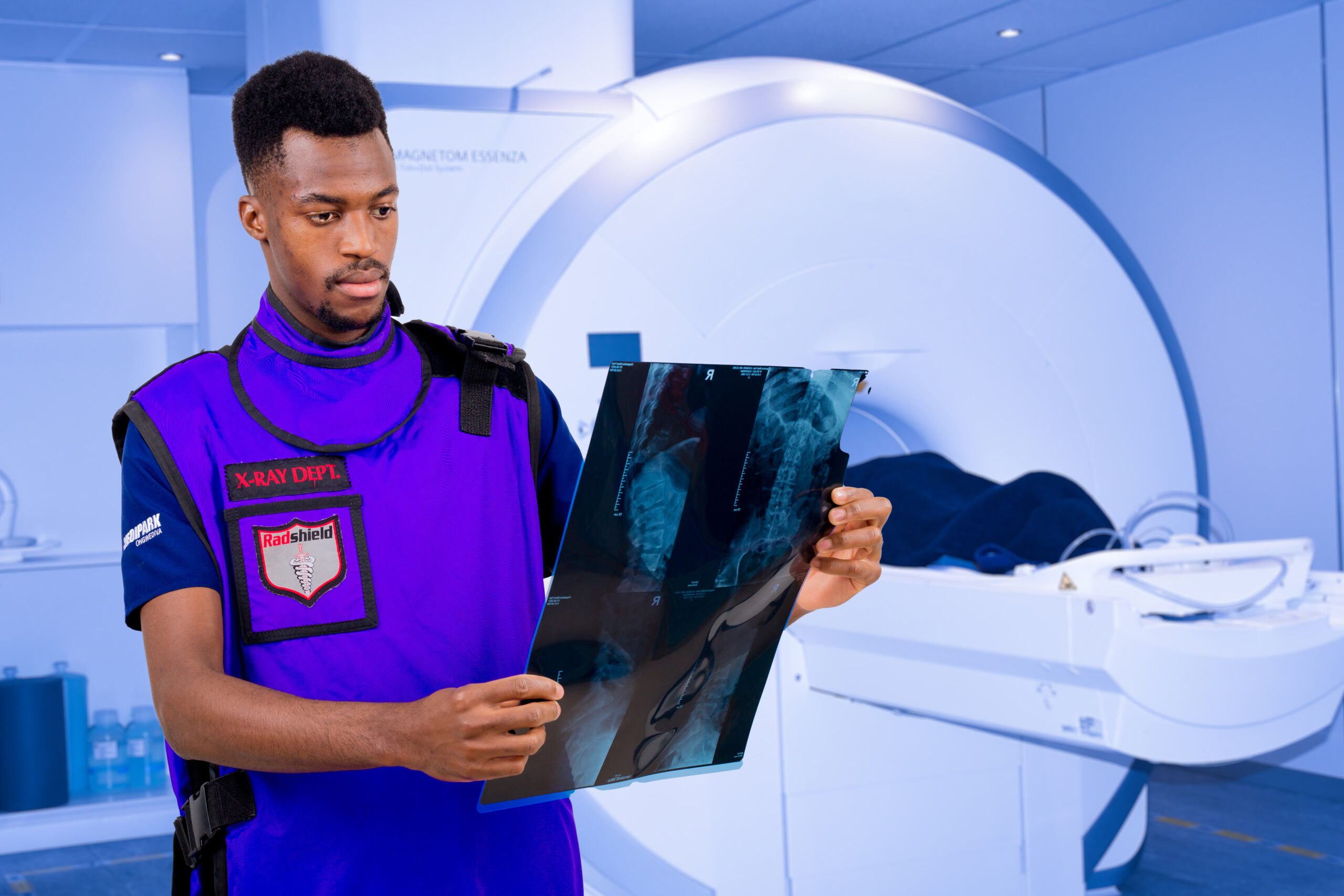
Welcome to Ongwediva Medipark Diagnostic Radiology, or simply OMDR. OMDR provides diagnostic and interventional radiology services and opened its doors in Nov 2006. From that auspicious beginning OMDR has since grown and currently offers most if not all imaging modalities.

OMDR is dedicated to providing the highest quality imaging and interventional services available, utilizing state of the art equipment, in conjunction with advanced, up-to-date techniques.
Our radiologists are certified Fellows of the College of Diagnostic Radiologists of South Africa. They are subspecialised in Neuroradiology, MSK, Interventional, breast imaging, HPB studies.
We are fully committed to provide excellence in service to both our patients and our referring medical doctors. It is our number one priority.
We offer the following services at OMDR:
Computed Tomography (CT Scan) is a computerized x-ray imaging procedure that produces cross-sectional images of the body which can be viewed in two or three dimensions. These scans are used for examination of the brain, heart, bone density, chest, and abdominal structures as well as other fine detail examinations of the spine and joints.
Duration of test: 10-30minutes
What to expect: You may be requested to change into a hospital gown. You will then be asked to lie on a table that slides into the scanner, which looks like a large doughnut. The x-ray rotates around you to take the images. If more details are requested, the radiographer may put up a cannula and inject a dye into your veins or give you a solution to take orally. Some of these examinations may require additional time.
MRI
Magnetic resonance imaging (MRI) is a medical imaging technique used in radiology to form pictures of the anatomy and the physiological processes of the body. MRI scanners use strong magnetic fields, magnetic field gradients, and radio waves to generate images of the organs in the body. Unlike CT scans or X-rays, MRI doesn’t use radiation.
Duration of test: 45minutes per region
What to expect: You will be asked to change into a hospital gown. Further screening questions will be conducted to ensure that you do not enter with metallic objects into the MRI room as this may be very dangerous for yourself or the radiographer. You will then be asked to lie on the table that slides into the MRI machine. The MRI magnet creates a loud tapping or thumping noise. Headphones will be given to you to cut out the loud noise and will also allow you to communicate to the radiographer. Some patients may feel claustrophobic because of the confined space. This discussion will take place between yourself and the radiographer who will answer all your questions and put you at ease. In some cases, a mild sedative may be given.
Fluoroscopy is an imaging technique that uses X-rays to obtain real-time moving images of the interior of a body part. Fluoroscopy is used in a wide variety of examinations and procedures to diagnose or treat patients. Some examples are: Barium X-rays and enemas (to view the gastrointestinal tract) Catheter insertion and manipulation (to direct the movement of a catheter through blood vessels, bile ducts or the urinary system)
Duration of test: 10minutes to more
For other interventional procedures like barium enema, barium swallow studies these may take up to more than 2hours.
What to expect: You may be requested to change into a hospital gown as your clothing may interfere with image quality. You will then be taken into the screening room and asked to lie, sit or stand while the x-ray machine takes images of the area under investigation. You may be moved into different position to get the images.
Mammography: Mammography is the process of using low-energy X-rays to examine the human breast for diagnosis and screening. The goal of mammography is the early detection of breast cancer, typically through detection of characteristic masses or microcalcifications.
Duration of test: 10min to an hour
In some cases, an ultrasound will be done before starting with the mammogram which may take additional time.
What to expect: You will be requested to change into a hospital gown. You will then be taken into the mammo room and asked to stand while the x-ray machine takes images of the breast. Expect a bit of sensitivity as your breast may be compressed to get high-quality detail. Inform the radiographer when your breast are too painful during the exam.
An ultrasound scan uses high-frequency sound waves to make an image of a person's internal body structures. Doctors commonly use ultrasound to study a developing fetus (unborn baby), a person's abdominal and pelvic organs, muscles and tendons, or their heart and blood vessels.
Duration of test: 30min to 1hour
What to expect: For some ultrasounds, you may be asked to change into a hospital gown. Then asked to sit or lie on the sonar room bed. The radiologist/sonographer will apply gel to the skin on area of investigation, then presses a small probe against it, moving it to capture images of the inside of the body. In some examinations of the abdomen and lower pelvis, you will be asked to drink water and wait for certain minutes before exam is done. Some other ultrasounds may require the use of a probe that goes into the vaginal or rectum. The reasons for this will be discussed with you by the performing technician before the exam.
Imaging with x-rays involves exposing a part of the body to a small dose of ionizing radiation to produce pictures of the inside of the body. ... X-rays are the oldest and most frequently used form of medical imaging. The most common plain film x-rays are chest x-rays used to mostly diagnose lung disease.
Duration of test: 10-15minutes
What to expect: You may be requested to change into a hospital gown as some of the clothing may interfere with image quality. You will then be taken into the x-ray room and asked to lie, sit or stand while the x-ray machine takes images of the area under investigation. You may be moved into different position to get the images.

Department Doctors

en_USEnglish